Latest Photo Galleries


Brazilian Markets
17h36 Bovespa |
-0,17% | 124.171 |
16h43 Gold |
0,00% | 117 |
16h59 Dollar |
-0,48% | 5,2424 |
16h30 Euro |
+0,49% | 2,65250 |
ADVERTISING
Brazilian Researchers Make 3D Neuron Image With Particle Accelerator
08/15/2018 - 14h12
Advertising
PHILLIPPE WATANABE
SÃO PAULO
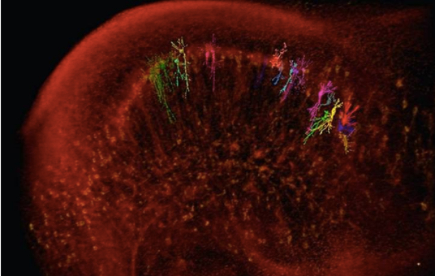
A neuron in 3D. This is what Brazilian scientists obtained after making X-ray microtomographies with a particle accelerator. This may improve the understanding of diseases such as Alzheimer's or Parkinson's.
The technique can be described as rotating a brain sample in from of a X-ray beam. Like in a puzzle, the resulting 2048 images are joined with the use of math and computers, forming a 3D image of brain and neurons.
One of the main advantages of this method is its ease. "We are able to get a cell image in its complete state," said Matheus Fonseca, researcher at LNBio (National Bioscences Laboratory), one of the paper's authors.
| divulgação | ||
 |
||
| A neuron in 3D |
Currently, the usual practice requires cleaning and slicing the brain that will be studied. But in the new methodology developed by the Brazilian researchers, all that is needed is to dip the organ in a mercury solution and do the tomography.
This is where the particle accelerator from LNLS (National Laboratory of Synchrothron Light), in Campinas (SP). The researchers used the accelerator's radiation to create the 3D images.
As soon as Sirius - a second fourth-generation source of synchrothron light (radiation produced with the particle acceleration process) – becomes active, the scientists hope to obtain resolution ten times greater than presently.
Translated by NATASHA MADOV